New Patients
(860) 218-9463
Existing Patients
(860) 421-0144

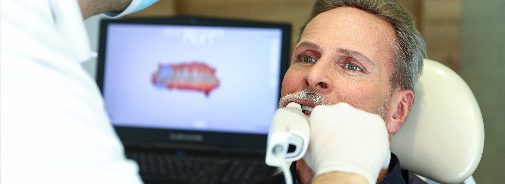
Digital impressions use a compact intraoral scanner to capture hundreds or thousands of precise images of your teeth and gums, which are stitched together by software to create a three-dimensional model. Unlike traditional putty impressions, the scanner records surface detail, occlusion and surrounding soft tissue in real time. That 3D model becomes the foundation for crowns, bridges, veneers, implant restorations and many other treatments, allowing clinicians to plan and communicate treatment with a degree of clarity that was not previously possible.
Because the output is a computerized dataset rather than a physical mold, clinicians can manipulate the model on a screen instantly: rotate it, zoom in on margins, and verify interproximal contacts and bite relationships. This immediacy helps identify any areas that need rescanning right away, reducing the likelihood of delays later in the case. For patients, the result is a faster diagnostic process with fewer surprises and more predictable restorative outcomes.
Scanners vary in design and capability, but all modern intraoral systems prioritize accuracy, ease of use and seamless integration with laboratory and CAD/CAM workflows. The quality of a digital impression depends on good technique and clear visualization, which experienced clinicians can obtain consistently during routine appointments. Ultimately, digital impressions are a tool that improves communication, efficiency and clinical precision across a wide range of dental procedures.
One of the most immediate benefits patients notice is comfort. Traditional impressions often require viscous materials and impression trays that can trigger gagging or create an unpleasant oral sensation. Digital scanning removes the need for those materials: the scanner’s wand is guided around the mouth while live images appear on a monitor. The procedure is typically quicker and less invasive, which makes it especially helpful for patients with strong gag reflexes, dental anxiety, or limited ability to tolerate long procedures.
Beyond physical comfort, digital impressions also enhance communication between the clinician and the patient. Because scans are visible in real time on the operatory screen, you can see what your dentist sees—margins, occlusal relationships and areas of wear or decay are easier to discuss when shown visually. This shared view helps patients understand recommended treatments and the rationale behind clinical decisions, supporting informed consent and collaborative treatment planning.
The convenience factor extends after the appointment as well. Digital files are easy to store, back up and retrieve for future reference. That means subsequent visits that require reference to a prior scan—such as follow-up restorative adjustments or replacement of a restoration—can proceed without repeating invasive steps, saving chair time and preserving patient comfort.
Precision is a core advantage of digital impressions. High-resolution scans capture fine anatomic details that influence how a restoration fits and functions—crown margins, pontic contours, and occlusal contacts are all represented in the digital model. When these details are captured accurately, laboratory technicians or in-office CAD/CAM software can design restorations that seat more predictably and require fewer adjustments at delivery.
Digital workflows also improve communication with dental laboratories by providing standardized, measurable files that technicians can use directly with CAD systems. The result is improved consistency from case to case and improved reproducibility for complex prosthetic work. Additionally, scans can help preserve soft-tissue contours in the digital record, which is important for esthetic cases and implant restorations that rely on precise tissue architecture.
From a clinical standpoint, fewer adjustments at try-in mean less time in the chair for patients and a lower chance of complications related to misfit. Accurate digital records also make it simpler to track changes over time, support shade selection when combined with high-quality photography, and facilitate multidisciplinary planning for cases that involve orthodontics, periodontics or implant therapy.
Once a scan is captured, the digital file becomes the central data point in a streamlined workflow. Files can be shared electronically with a dental laboratory or used in the office with milling systems to fabricate restorations in a single visit. That flexibility allows clinicians to choose the best pathway for each patient—outsourced laboratory fabrication for complex cases or in-office milling for expedient single-unit restorations when appropriate.
Electronic transmission of files reduces logistical steps associated with shipping physical impressions and stone models. Laboratories receive standardized data quickly, which shortens the administrative handoff and helps coordinate scheduling and case planning. For practices that collaborate with multiple labs, consistent digital files reduce variability and support more efficient lab feedback loops.
Digital models also support advanced treatment planning tools, such as virtual articulation, implant surgical guides and digital smile design. These capabilities empower team-based planning and can improve the predictability of multi-disciplinary treatment. Because the data are digital from the outset, clinicians have the freedom to revisit the case, make iterative changes and archive versions of the model for legal and clinical records without occupying physical storage space.
At our Unionville office on Farmington Ave, we integrate intraoral scanning into everyday care to improve outcomes and patient experience. Our clinical team is trained to use the latest scanners in a way that complements conservative treatment planning, accurate restorations and clear patient communication. The equipment is maintained and calibrated according to manufacturer guidelines to preserve accuracy across all types of restorative and implant cases.
Implementing digital impressions also supports infection control and recordkeeping. Because impressions are captured digitally, there is less handling of impression materials and fewer items that require disinfection or storage. Digital files become part of the patient record, simplifying follow-up care and making it easier to coordinate with specialists or partner laboratories. This digital-first approach is designed to support continuity of care across appointments and practitioners.
For patients preparing for a scan, little special preparation is needed—good oral hygiene and a short pre-procedure rinse are typically sufficient. During the appointment the clinician will explain the process and review images together so you understand what’s being recorded and why. If a case requires laboratory collaboration or in-office milling, we will outline the anticipated workflow so you know what to expect at each stage.
In summary, digital impressions are a modern, patient-friendly way to capture the detail clinicians need for precise, predictable dentistry. They improve comfort, enhance communication, and support efficient workflows that benefit both patients and providers. To learn more about how digital scanning may be used in your care at Newpoint Family Dental or to discuss whether it’s right for an upcoming procedure, please contact us for more information.
