New Patients
(860) 218-9463
Existing Patients
(860) 421-0144


An intraoral camera is a compact, wand-like imaging device designed specifically for use inside the mouth. Unlike a typical camera, it is built to capture clear, full-color images of teeth, gums, and other soft tissues from angles that are difficult to view with the naked eye. These images appear almost immediately on a monitor, giving both clinician and patient a detailed visual reference during an exam.
Because the camera produces high-resolution stills and video, clinicians can document areas of concern with precision. This capability makes small cracks, early decay, worn tooth surfaces, and inflamed tissue visible in a way that routine visual inspection sometimes cannot achieve. The output is both diagnostic and educational—helping to bridge the gap between clinical findings and patient understanding.
Designed for comfort and efficiency, intraoral cameras are small enough to be maneuvered around the mouth with minimal disruption. They are typically used alongside other diagnostic tools, not as a replacement, providing an extra layer of visual information that supports more informed decision-making about care.
High-quality intraoral images support more accurate diagnosis by highlighting subtle changes in enamel, restorations, and soft tissue. When a suspected problem is visible on-screen, clinicians can evaluate the extent and nature of the issue and determine whether further testing—such as radiographs or direct clinical probing—is warranted. The images also help prioritize treatment needs based on what is seen rather than relying solely on surface-level symptoms.
Images captured with an intraoral camera integrate well into digital patient records, allowing clinicians to compare current photos with prior visits. This chronological view makes it easier to track progression or healing over time, which is especially helpful for monitoring early decay, the integrity of fillings and crowns, and periodontal changes.
Because intraoral imaging can be reviewed with the patient in real time, it supports collaborative treatment planning. Visual evidence helps patients understand why a recommendation is being made, which can improve acceptance of necessary care and foster clearer communication about alternatives, timelines, and follow-up steps.
One of the most tangible benefits of intraoral imaging is its ability to make the invisible visible. When patients see close-up images of their own teeth and gums, complex findings become straightforward to explain. This transparency builds trust and helps patients feel more engaged in their care.
Images can be annotated during an appointment to point out specific concerns, such as the margins of an old restoration, a hairline fracture, or localized gum irritation. These marked images serve as a lasting visual record that patients can reference afterward, making it easier to recall the clinician’s recommendations and the reasons behind them.
For cases that require coordination with specialists or a dental laboratory, intraoral photos can be shared as part of a clear, documented case file. These visuals help outside providers understand the situation quickly and can improve the consistency of care across multiple professionals without relying solely on written descriptions.
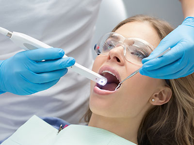
Using an intraoral camera is quick and noninvasive. During a routine exam or a focused visit, the clinician will gently guide the camera around the mouth while you sit comfortably. The process usually takes only a few minutes and is suited to most patients, including adults and older children who can hold still for brief periods.
The camera’s live feed allows the clinician to explain what is being seen as it happens. In many examinations, photos are taken for documentation—these images become part of the patient’s chart and can be revisited at future appointments. The streamlined workflow helps save time and reduces the need for repeated chairside explanations.
Because the device is used intraorally, strict infection-control protocols are followed. Single-use sleeves or sterilizable barriers and routine disinfection of external surfaces ensure the process adheres to standard safety guidelines and protects patient health.
Intraoral cameras form a useful component of a modern, digital dental practice. Their images can be stored in electronic health records, linked with digital radiographs, or included in treatment planning software. This interoperability improves clinicians’ ability to form comprehensive diagnoses and creates a centralized repository of visual data for each patient.
Advanced systems now offer features such as image enhancement, measurement tools, and the ability to export standardized files for collaboration with dental specialists or laboratories. These enhancements speed communication and reduce the likelihood of misinterpretation when treatment is coordinated across providers.
As imaging technology evolves, intraoral cameras continue to gain resolution and functionality while remaining simple to use. The trend toward integrated digital workflows—combining intraoral photography, digital impressions, and radiography—supports more predictable outcomes and more efficient care delivery.
In summary, intraoral cameras are a practical, patient-friendly tool that enhances diagnosis, documentation, and communication during dental care. By making detailed images available in real time, this technology helps patients better understand their oral health and supports clinicians in planning effective treatment. If you’d like to learn more about how we use intraoral imaging in our Unionville practice, please contact us for additional information and to discuss how these tools can benefit your care.